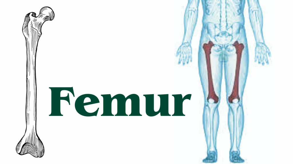
Understanding Femur Health
The femur, commonly referred to as the thigh bone, holds the distinction of being both the longest and strongest bone in the human body. Its remarkable length and robust structure play a crucial role in the skeletal system. Extending from the hip to the knee, the femur is integral in supporting the body’s weight and enabling a wide range of movements.
One of the primary functions of the femur is to act as a pillar of support. Given its position and structural integrity, it bears the brunt of the body’s weight, particularly when standing, walking, or running. This bone’s strength is vital for maintaining balance and ensuring that the body can perform everyday activities with ease.
Beyond its support role, the femur is essential for mobility. It is intricately involved in various movements such as walking, running, and jumping. The femur’s articulation with the hip and knee joints allows for a wide range of motion, providing the flexibility and strength necessary for these activities. The bone’s unique structure, with a slightly curved shape and a robust shaft, contributes to its ability to withstand significant stress and impact.
The femur’s importance extends beyond just physical support and movement. It also houses bone marrow, which is crucial for producing blood cells. This multi-functional nature highlights the femur’s critical role in both the musculoskeletal and hematopoietic systems.
Understanding the femur’s anatomy and function provides insight into its significance in the human body. Its extraordinary strength, coupled with its essential role in movement and support, underscores why it is often regarded as a cornerstone of the skeletal system. As we delve deeper into the specifics of the femur, its fascinating attributes and contributions to human health and mobility become even more apparent.
Anatomy of the Femur
The femur, commonly known as the thigh bone, is the longest and strongest bone in the human body. It plays a crucial role in supporting the weight of the body and facilitating movement. The femur is divided into several key parts: the head, neck, shaft, and distal end, each with distinct anatomical features and functions.
The head of the femur is a spherical structure that articulates with the acetabulum of the pelvis, forming the hip joint. This ball-and-socket joint allows for a wide range of motion, including flexion, extension, abduction, and rotation. The head is covered with articular cartilage, which reduces friction and absorbs shock during movement.
Connecting the head to the shaft is the neck of the femur. This narrow, cylindrical segment is angled to optimize the alignment of the hip joint and ensure efficient weight distribution. The neck is susceptible to fractures, particularly in older adults due to osteoporosis or trauma.
The shaft, also referred to as the diaphysis, constitutes the longest portion of the femur. It has a slight anterior bow and is composed of dense, compact bone that provides structural integrity. The shaft serves as an anchor point for various muscles, including the quadriceps, adductors, and hamstrings, which are essential for leg movement and stability.
The distal end of the femur widens to form the medial and lateral condyles, which articulate with the tibia and patella to form the knee joint. The condyles are covered with articular cartilage, facilitating smooth and pain-free motion. Additionally, the femur features several bony prominences, such as the greater and lesser trochanters, which serve as attachment sites for ligaments, tendons, and muscles.
In summary, the femur’s complex anatomy is integral to its function in the human body. Its various parts, including the head, neck, shaft, and distal end, work together to support movement and stability, making it a vital component of the musculoskeletal system.
Functions of the Femur
The femur, as the longest and strongest bone in the human body, plays a pivotal role in various physiological functions. Primarily, the femur is essential for weight-bearing. It supports the body’s weight during both static postures, such as standing, and dynamic activities, including walking and running. This remarkable bone is capable of withstanding immense pressure, ensuring stability and balance.
Another critical function of the femur is its role in facilitating movement. The femur acts as a lever that allows for a wide range of motions, thanks to its articulation with the hip and knee joints. This bone’s structural integrity and connection to these joints enable complex movements such as flexion, extension, abduction, and rotation, which are fundamental for various physical activities. These movements are crucial for daily functions, athletic endeavors, and overall mobility.
Additionally, the femur serves as a significant site for muscle attachment. Numerous muscles of the thigh and hip region, including the quadriceps, hamstrings, and gluteal muscles, attach to the femur. These muscles play a vital role in movement and stability. When these muscles contract, they exert force on the femur, generating the necessary movements for locomotion and other physical activities. This interconnectedness highlights the femur’s importance in overall muscular function and coordination.
Furthermore, the femur contributes to maintaining balance and stability in the human body. The bone’s length and position help in distributing the body’s weight evenly across the lower limbs. This distribution is crucial for maintaining an upright posture and preventing falls. The femur’s ability to support and distribute weight effectively ensures that the body’s center of gravity is maintained, facilitating balance during both movement and stationary positions.
In essence, the femur’s multifaceted functions underscore its significance in the human skeletal system. Its role in weight-bearing, movement facilitation, muscle attachment, and balance support highlights the femur’s indispensable contribution to the body’s overall functionality and stability.

Common Femur Injuries and Conditions
The femur, being the longest and one of the strongest bones in the human body, is crucial for supporting our weight and enabling mobility. Despite its robustness, the femur is susceptible to various injuries and medical conditions. Understanding these common issues can aid in prompt diagnosis and effective treatment.
Fractures are among the most frequent femur injuries. High-impact trauma, such as car accidents or falls from significant heights, often leads to femoral fractures. Symptoms typically include severe pain, inability to bear weight, and visible deformity. Treatment usually involves surgical intervention, such as internal fixation or the insertion of rods and screws, followed by rehabilitation to restore full function.
Osteoporosis is another condition that significantly affects the femur. This systemic disease results in decreased bone density and increased fragility, making the femur more prone to fractures even with minor falls or stresses. Postmenopausal women and the elderly are particularly at risk. Management of osteoporosis includes medication to strengthen bones, dietary adjustments to ensure adequate calcium and vitamin D intake, and weight-bearing exercises to improve bone health.
Femoral head necrosis, also known as avascular necrosis, is a condition where the blood supply to the femoral head is compromised, leading to bone tissue death. This condition can result from trauma, prolonged steroid use, or excessive alcohol consumption. Symptoms include pain in the hip or groin area, stiffness, and reduced range of motion. Treatment varies based on the stage of the condition but may include medication, physical therapy, or surgical options like core decompression or hip replacement.
Overall, awareness of these common femur injuries and conditions is essential for early detection and appropriate intervention. Regular check-ups, maintaining a healthy lifestyle, and adhering to safety measures can significantly reduce the risk of femur-related complications.
Diagnosis and Treatment of Femur Problems
Diagnosis of femur-related issues typically begins with a comprehensive physical examination, during which a healthcare provider assesses the patient’s medical history and current symptoms. To confirm the diagnosis, imaging techniques such as X-rays, Magnetic Resonance Imaging (MRI), and Computed Tomography (CT) scans are frequently employed. X-rays are particularly effective in identifying fractures, dislocations, and bone deformities. MRI and CT scans provide more detailed images, allowing for the evaluation of soft tissues, ligaments, and the extent of bone damage.
Once a diagnosis is established, the treatment approach varies based on the specific condition and its severity. Non-surgical treatment options often include immobilization techniques, such as casting or bracing, to stabilize the bone and facilitate natural healing. Additionally, pain management strategies, including medications and activity modification, are implemented to enhance patient comfort during the recovery process.
In cases where non-surgical methods are insufficient, surgical intervention may be required. Common surgical procedures include internal fixation, where metal rods, screws, or plates are used to hold the bone fragments in place, and hip replacement surgery for severe cases involving the femoral head. These procedures aim to restore the structural integrity of the femur and enable proper functionality.
Post-surgery, physical therapy and rehabilitation are crucial components of the recovery journey. Tailored exercise programs designed by physical therapists help in regaining strength, flexibility, and mobility. Regular follow-up appointments are essential to monitor progress and adjust the rehabilitation plan as needed.
Ultimately, the combination of accurate diagnosis and appropriate treatment ensures optimal outcomes for individuals experiencing femur problems. By understanding the diagnostic procedures and treatment options available, patients can navigate their recovery with confidence and achieve the best possible results.
Maintaining Femur Health
The femur, known as the longest bone in the human body, plays a crucial role in our overall mobility and stability. Maintaining femur health is essential for ensuring long-term bone strength and functionality. A multifaceted approach that includes proper nutrition, targeted exercises, and lifestyle modifications can significantly contribute to the well-being of this vital bone.
Proper nutrition is foundational for femur health. A diet rich in calcium and vitamin D is crucial as these nutrients are vital for bone density and strength. Dairy products, leafy greens, and fortified foods are excellent sources of calcium, while exposure to sunlight and consumption of fatty fish and fortified dairy products can help maintain adequate levels of vitamin D. Additionally, incorporating foods high in magnesium, such as nuts and seeds, and phosphorus-rich foods like meat and poultry, can further support bone health.
Regular physical activity is another key element in maintaining a healthy femur. Weight-bearing exercises, such as walking, running, and resistance training, can help strengthen the femur and surrounding muscles. Incorporating exercises that target the lower body, like squats and lunges, can enhance muscle support around the femur, reducing the risk of fractures and injuries. Balance and flexibility exercises, such as yoga and pilates, can also improve overall stability and reduce the likelihood of falls.
Lifestyle changes are equally important in preventing femur-related injuries and conditions. Avoiding smoking and excessive alcohol consumption is beneficial, as both can negatively impact bone density. Ensuring a healthy weight is another critical factor, as being either underweight or overweight can put undue stress on the femur. Wearing appropriate footwear and practicing good posture can also mitigate unnecessary strain on the lower body.
By adopting a comprehensive approach that includes proper nutrition, regular exercise, and mindful lifestyle choices, individuals can effectively maintain the health of their femur. These strategies not only promote bone strength but also enhance overall physical well-being, contributing to a healthier, more active life.

The Femur in Different Life Stages
The femur, known as the thigh bone, undergoes significant transformations throughout various stages of life. During childhood and adolescence, the femur experiences rapid growth, especially noticeable during growth spurts. This period is characterized by the elongation of the bone, driven by the activity in the growth plates located at its ends. These growth plates, or epiphyseal plates, are areas of cartilage that gradually ossify and turn into bone as the individual matures. Proper nutrition, physical activity, and overall health are critical during this phase to ensure optimal femur development.
As individuals transition into adulthood, the femur reaches its maximum length and density. The bone’s structure becomes more robust and capable of supporting increased physical demands. The adult femur is integral to mobility and weight-bearing activities, providing a crucial connection between the hip and knee joints. Maintaining femur health during adulthood is essential, as it plays a significant role in an individual’s overall mobility and quality of life. Regular exercise, a balanced diet rich in calcium and vitamin D, and avoiding excessive strain are pivotal in preserving femur strength and function.
In the later stages of life, the femur can undergo various age-related changes. Bone density may decrease due to a reduction in bone mass, a condition known as osteoporosis, which can make the femur more susceptible to fractures. This is particularly concerning for the elderly, as femur fractures can lead to severe complications and prolonged recovery periods. Preventive measures such as routine bone density screenings, weight-bearing exercises, and adequate nutritional intake become increasingly important to mitigate these risks.
Throughout all life stages, the health of the femur is influenced by a combination of genetic, lifestyle, and environmental factors. Understanding these changes and implementing strategies to support femur health can enhance overall well-being and mobility across the lifespan.
FAQ About the Femur
What is the femur?
The femur, also known as the thigh bone, is the longest and strongest bone in the human body. It extends from the hip to the knee and plays a crucial role in supporting the weight of the body, enabling movement, and maintaining posture.
How long does it take for a femur to heal?
The healing time for a femur fracture can vary widely depending on the severity of the break, the patient’s age, and overall health. On average, it takes about 3 to 6 months for a femur to heal completely. However, some complex fractures may require a longer recovery period, and rehabilitation exercises are often necessary to restore full function.
What are common symptoms of a femur fracture?
Common symptoms of a femur fracture include severe pain, an inability to bear weight on the affected leg, swelling, bruising, and visible deformity or shortening of the leg. In some cases, the broken bone may be visible through the skin in an open fracture.
Can you walk with a broken femur?
Walking with a broken femur is typically not possible due to the intense pain and instability of the leg. Immediate medical attention is required to properly align the bone and facilitate healing. Patients usually need crutches or a wheelchair during the initial recovery phase and may gradually progress to walking with the help of physical therapy.
Can a femur fracture heal without surgery?
In some cases, a femur fracture can heal without surgery, especially if the break is stable and the bones are properly aligned. Non-surgical treatment typically involves immobilization with a cast or brace. However, if the fracture is displaced or complex, surgery may be necessary to realign the bones and ensure proper healing.
What is the recovery process after femur surgery?
The recovery process after femur surgery involves a combination of rest, pain management, and physical therapy. Initially, weight-bearing may be limited, and the patient may need assistive devices like crutches or a walker. Over time, physical therapy will help strengthen the muscles and improve mobility. Full recovery can take several months, depending on the severity of the fracture and the patient’s progress.
Can a femur fracture cause long-term complications?
While most femur fractures heal with no long-term problems, complications can occur, especially with severe fractures or improper healing. These may include chronic pain, arthritis in the hip or knee joints, muscle weakness, or limited range of motion. In rare cases, improper alignment during healing may require further surgery to correct.
These answers aim to provide a basic understanding of the femur and the implications of a femur fracture. For more detailed information or personalized advice, consulting a medical professional is always recommended.

Discover more from HUMANITYUAPD
Subscribe to get the latest posts sent to your email.

