Photo 51: The Shocking Truth Behind DNA Discovery

Understanding Photo 51
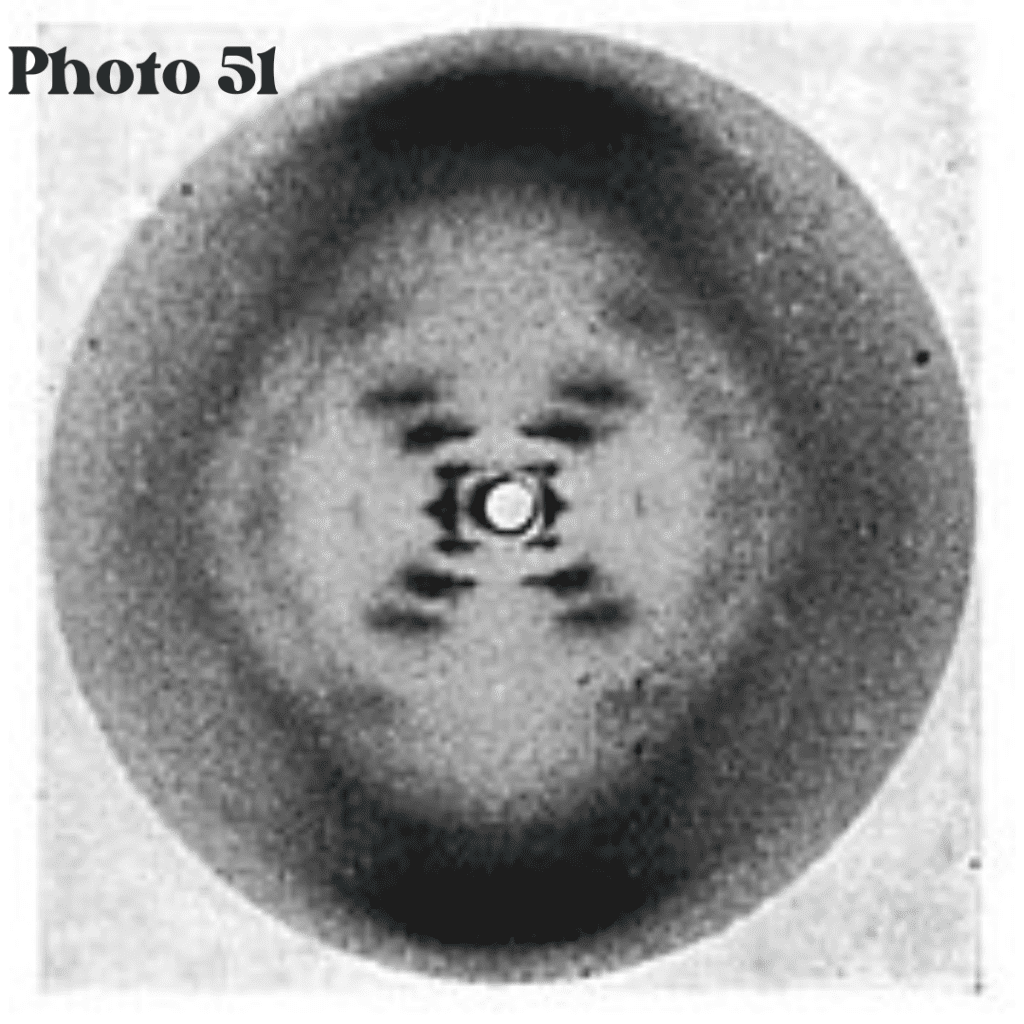
Photo 51 is a seminal X-ray diffraction image that played a critical role in unraveling the structure of DNA, fundamentally altering our understanding of genetics. Captured in 1952 by Rosalind Franklin and her team at King’s College London, this image served as a crucial piece of evidence in establishing the double helix model of DNA. The creation of Photo 51 occurred during an era characterized by intense research into the molecular biology of cells, and the mystery surrounding genetic material was a primary focus of scientific inquiry.
In the early 1950s, the quest to uncover the structure of DNA was underway, with various scientists competing to elucidate the genetic blueprint of life. Franklin, a skilled crystallographer, utilized X-ray diffraction techniques to obtain images that revealed the helical structure of DNA. Photo 51, in particular, provided a distinct diffraction pattern that suggested a helical organization, indicating that DNA consisted of two intertwined strands. This finding was pivotal not only for molecular biology but for the broader field of genetics, linking the physical structure of DNA to its biological function in encoding genetic information.
The significance of Photo 51 extends beyond its technical contribution; it also underscores the collaborative, yet competitive nature of scientific research during this period. The eventual interpretation of Photo 51 by James Watson and Francis Crick, who integrated Franklin’s findings with their own research, culminated in the proposal of the double helix model for DNA. This groundbreaking moment set the stage for modern genetics, providing insights that would lead to advancements in molecular biology, medicine, and biotechnology. Overall, Photo 51 remains a symbol of the intersection between technology and discovery in the scientific narrative of DNA.
The Historical Background of DNA Research
The journey of DNA research has evolved significantly since its early contemplation in the latter part of the 19th century. Initially, the building blocks of heredity were obscure, and the scientific community largely perceived traits as resultant phenomena without a molecular basis. The field began to take shape with the foundational work of Gregor Mendel in the 1860s, who established the concept of inheritance through his meticulous pea plant experiments. Mendel’s laws of inheritance set the groundwork for future genetic exploration, albeit his findings were largely overlooked until the early 20th century.
In the 1900s, the understanding of chromosomes and their role in heredity began to emerge. Pivotal figures such as Thomas Hunt Morgan and his fruit fly experiments revealed the significance of chromosomes in genetic traits, paving the way for the chromosomal theory of inheritance. This marked a crucial shift towards molecular biology, where scientists started to manipulate and delve into the genetic material itself.
By the 1940s, the field entered a new paradigm with the discovery that nucleic acids, namely DNA, served as carriers of genetic information. The work of scientists like Erwin Chargaff contributed to understanding the composition of DNA. Chargaff’s rules, which explained the consistent ratios of adenine to thymine and cytosine to guanine, indicated a potential structure of DNA, yet the three-dimensional structure remained elusive.
Overall, the efforts of these researchers laid the groundwork for a deeper comprehension of molecular genetics. Their contributions, combined with advancements in technology and the burgeoning field of biochemistry, created an environment ripe for the groundbreaking discovery of Photo 51. This pivotal moment would ultimately illuminate the double helical structure of DNA and revolutionize biological sciences, leading to numerous breakthroughs in genetics and molecular biology.
The Science Behind X-ray Diffraction
X-ray diffraction (XRD) is a powerful analytical technique utilized to determine the atomic and molecular structure of a crystal. It involves directing X-rays onto a crystalline sample and analyzing the resulting scattering pattern. When X-rays penetrate a crystal, they are scattered by the electron clouds surrounding the atoms within the crystal lattice. This scattering produces a unique pattern of diffraction, which can be recorded on a detector. The arrangement of atoms in the crystal affects the angles and intensities of the scattered X-rays, enabling researchers to deduce information about the atomic structure.
The fundamental principle of X-ray diffraction lies in Bragg’s Law, which states that constructive interference occurs when X-rays reflect off the crystal planes at specific angles. This law can be mathematically expressed as nλ = 2dsinθ, where n is an integer, λ is the wavelength of the X-rays, d is the distance between crystal planes, and θ is the angle of incidence. By varying the angle and measuring the intensity of the diffracted beams, scientists can derive a three-dimensional picture of the electron density within the crystal, leading to the identification of atomic positions.
This method has proven particularly effective for studying biological macromolecules such as DNA due to their crystalline nature. Crystallization of DNA allows for a well-ordered arrangement of molecules, creating a suitable environment for X-ray diffraction analysis. The resulting diffraction patterns can reveal vital information about the molecular structure, including helical features, spacing between base pairs, and overall conformational arrangements. In the case of Photo 51, the X-ray diffraction technique played a crucial role in elucidating the double helix structure of DNA, paving the way for groundbreaking discoveries in molecular biology and genetics.

Rosalind Franklin’s Contribution
Rosalind Franklin was an eminent scientist whose work in the field of X-ray crystallography was pivotal to understanding the molecular structure of DNA. Born in 1920 in London, Franklin pursued her education in physical chemistry at the University of Cambridge, where her exceptional analytical skills emerged. Her expertise in techniques for studying crystallized substances enabled her to capture the now-famous Photo 51 in 1952, a remarkable X-ray diffraction image of DNA. This image provided crucial insights into the helical structure of DNA, yet Franklin’s contributions were often overshadowed by her male counterparts.
Franklin’s methodology was characterized by meticulous attention to detail and stringent experimental rigour. She employed advanced X-ray techniques to analyze samples of DNA, successfully producing high-quality images despite the challenges posed by the complex molecular structure. Her systematic approach allowed her to deduce vital information about DNA, including its dimensions and the orientation of its phosphate backbone. This dexterity in handling intricate tools demonstrated her profound scientific acumen, positioning her as a leading figure in this domain.
However, her journey was fraught with challenges, particularly as a woman in a predominantly male scientific community. Franklin faced significant gender bias, which contributed to her work being undervalued and often misattributed. While James Watson and Francis Crick capitalized on her findings to propose the double-helix model of DNA, Franklin continued to pursue her own research, unyielding in the face of adversity. Her analysis of the diffraction patterns of DNA was vital in corroborating the helical structure, making her an essential pioneer in the field of genetics. In light of her steadfast efforts and groundbreaking insights, Rosalind Franklin’s contribution to DNA discovery remains an integral part of scientific history.
The Reaction of Watson and Crick
In the early 1950s, James Watson and Francis Crick were engaged in an intense pursuit to unravel the structure of deoxyribonucleic acid (DNA), a goal that had eluded many scientists before them. Their groundbreaking discovery was significantly influenced by the X-ray diffraction image known as Photo 51, produced by Rosalind Franklin. Upon encountering this image, Watson and Crick experienced a profound moment of realization regarding the molecular configuration of DNA.
The distinct “X” pattern observed in Photo 51 provided Watson and Crick with crucial insights into the helical structure of DNA. They interpreted the symmetry and spacing of the diffraction patterns as indicative of a double helix, a structure composed of two intertwined strands. This image served as the catalyst for their theoretical framework, leading them to postulate that DNA consists of two sugar-phosphate backbones with bases pairing in the interior. Their interpretation was not only a pivotal moment in their research but also laid the foundation for molecular biology as a whole.
Watson and Crick’s collaborative efforts, informed by the pivotal information from Photo 51, underscored the importance of teamwork and shared scientific knowledge in the field of genetic research. The image illuminated aspects of DNA’s structure that had remained obscure, allowing them to envision a model that resonated with a broad range of biological functions, from replication to genetic encoding. The impact of their proposal was immediate and far-reaching, transforming the understanding of heredity and genetics.
Ultimately, the reaction to Photo 51 catalyzed a revolutionary shift in molecular biology, securing Watson and Crick’s place in scientific history. Their ability to appropriately leverage the insights gained from this critical X-ray image enabled them to elucidate the structure of DNA, revolutionizing the field and influencing generations of research thereafter.
The Implications of the Double Helix Model
The discovery of the double helix structure of DNA, elucidated through Photo 51, has had profound implications across various scientific disciplines, particularly genetics, molecular biology, and biochemistry. This groundbreaking model transformed our understanding of heredity and the mechanisms of genetic inheritance. Prior to this discovery, the precise molecular basis of heredity remained unclear, but the double helix provided a clear framework for understanding how genetic information is stored, replicated, and transmitted across generations.
The structure of DNA as a double helix suggests a methodical approach to genetic replication, where each strand of the helix serves as a template for creating a new complementary strand. This insight laid the foundation for understanding mutations—a change or alteration in the DNA sequence. The double helix model enables scientists to explore how mutations occur, their potential effects on organisms, and their role in the evolutionary process.
In the realm of molecular biology, the double helix model has been pivotal in guiding research on gene expression, regulation, and protein synthesis. The interactions between DNA, RNA, and proteins are essential for understanding cellular functions, leading to advances in biotechnology and genetic engineering. For instance, the ability to manipulate DNA through technologies such as CRISPR-Cas9 derives largely from the understanding gained through the double helix model.
Moreover, in biochemistry, the implications of the double helix extend to comprehending the biochemical pathways that govern cellular processes. The understanding of molecular interactions and the behavior of biomolecules in various environments has propelled research in pharmaceuticals, diagnostics, and therapeutic strategies, showcasing the transformative power of the double helix model. As our genetic comprehension expands, the double helix remains central to the ongoing exploration of life’s molecular basis and the complexities of biological systems.

Legacy of Photo 51 in Modern Science
Photo 51, taken by Rosalind Franklin in 1952, has left an indelible mark on modern science, particularly in the fields of molecular biology and genetics. This iconic X-ray diffraction image provided the first clear evidence of the helical structure of deoxyribonucleic acid (DNA), fundamentally transforming our understanding of genetic material. The implications of this discovery extend far beyond its initial revelation, influencing numerous biotechnological advances and genetic engineering practices today.
The clarity and precision of Photo 51 illuminated the path towards various innovations in genetic manipulation, such as CRISPR technology. By understanding the structure of DNA, researchers have been able to develop methods to edit genes with unprecedented accuracy. This capability paves the way for therapeutic interventions in genetic disorders, ushering in a new era of medicine. Techniques derived from insights gained from Photo 51 facilitate the study of gene functions and interactions, thereby enhancing our capacity to design targeted treatments that were previously unimaginable.
Moreover, the legacy of Photo 51 reverberates in forensic science, where DNA evidence plays a crucial role in criminal investigations. The methodologies established from the initial understanding of DNA structure enable forensic scientists to analyze genetic material effectively, leading to crucial breakthroughs in solving cases and exonerating the wrongly accused. The profound impact of Photo 51 is also evident in evolutionary biology. It has spurred studies on the genetic relationships among species, enhancing our comprehension of evolutionary processes and biodiversity.
In essence, the influence of Photo 51 is enduring, continuing to foster research and innovation across various scientific disciplines. This single image serves as a reminder of the intricate dance between discovery and technological advancement, inspiring countless scientists to explore the mysteries of life at the molecular level.
Ethical Considerations and Recognition
The advancements in scientific research, particularly in the study of DNA, exemplified by the discovery behind Photo 51, raise significant ethical considerations, particularly concerning credit and recognition. Central to this discussion is the role of Rosalind Franklin, whose meticulous work and pioneering efforts were pivotal, yet often inadequately acknowledged in the broader scientific narrative. Franklin’s contributions were instrumental in elucidating the structure of DNA, and her X-ray diffraction images provided critical insights that shaped subsequent research.
One of the notable ethical implications in this context is the challenge of ensuring appropriate recognition for all contributors in collaborative scientific endeavors. The case of Photo 51 highlights how the dynamics of scientific discovery can sometimes prioritize certain figures over others, leading to the overshadowing of key contributors like Franklin. This neglect raises questions about the integrity of scientific practice and the ethical responsibility of researchers to acknowledge all voices in collaborative settings.
Furthermore, the historical narrative surrounding the discovery of DNA often emphasizes the roles of James Watson and Francis Crick while neglecting to give due credit to Franklin. This discrepancy not only highlights a need for a more equitable approach to recognizing contributions in scientific work but also underscores the significance of transparency and inclusivity in research practices. Ethical considerations extend beyond mere acknowledgment; they are foundational to fostering a scientific culture that values diverse contributions and uplifts those who have been historically marginalized.
As the scientific community reflects on the legacy of Photo 51, it serves as a critical reminder of the importance of equitable recognition. By striving for an ethical framework that honors all contributors, the integrity of scientific research is fortified, paving the way for a more inclusive and responsible approach to discovery in the future.
Frequently Asked Questions about Photo 51
1. What is Photo 51?
Photo 51 is an X-ray diffraction image of DNA captured in 1952 by Rosalind Franklin and her team at King’s College London. It provided crucial evidence for the discovery of the double helix structure of DNA.
2. Why is Photo 51 important?
Photo 51 was instrumental in confirming that DNA has a helical structure. It helped James Watson and Francis Crick develop their double helix model, which became the foundation of modern molecular biology and genetics.
3. How was Photo 51 created?
Rosalind Franklin used X-ray diffraction techniques to analyze crystallized DNA fibers. The resulting diffraction pattern provided key structural details about DNA.
4. Who used Photo 51 to determine DNA’s structure?
James Watson and Francis Crick used insights from Photo 51—without Franklin’s direct permission—to finalize their double helix model of DNA in 1953.
5. Did Rosalind Franklin receive credit for Photo 51?
During her lifetime, Franklin’s contributions were largely overlooked. The 1962 Nobel Prize for the discovery of DNA’s structure was awarded to Watson, Crick, and Maurice Wilkins, omitting Franklin, who had passed away in 1958.
6. What is X-ray diffraction, and why is it useful for studying DNA?
X-ray diffraction is a technique used to determine the structure of crystalline substances by analyzing the way X-rays scatter off a sample. This method helped reveal DNA’s helical shape and precise molecular arrangement.
7. What did Photo 51 reveal about DNA’s structure?
Photo 51 showed a clear X-shaped diffraction pattern, indicating a helical structure with repeating elements, confirming that DNA consists of two intertwined strands.
8. What role did Maurice Wilkins play in Photo 51?
Maurice Wilkins, Franklin’s colleague at King’s College, showed Photo 51 to Watson and Crick without Franklin’s direct approval, enabling them to refine their DNA model.
9. How did Photo 51 contribute to modern science?
The discovery of DNA’s double helix has led to major advancements in genetics, biotechnology, medicine, forensic science, and genetic engineering.
10. Where is Photo 51 now?
Photo 51 is preserved at King’s College London, where it remains an important historical artifact in the history of molecular biology.

Discover more from HUMANITYUAPD
Subscribe to get the latest posts sent to your email.

