Unraveling the Symphony: The Vestibulocochlear Nerve’s Role in Human Perception

The human nervous system is a marvel of complexity, orchestrating a symphony of signals that govern our every movement and sensation. Among its many intricate components, the vestibulocochlear nerve stands out as a vital player in our ability to perceive and interact with the world around us. This cranial nerve, designated as the eighth cranial nerve, is a fascinating gateway that connects our auditory and vestibular systems. In this blog post, we will delve into the wonders of the vestibulocochlear nerve, exploring its anatomy, functions, and the crucial role it plays in our daily lives.
Anatomy of the Vestibulocochlear Nerve
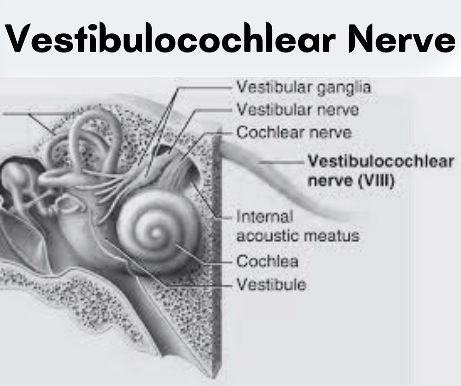
The vestibulocochlear nerve, also known as the eighth cranial nerve or simply CN VIII, is a paired nerve that consists of two components: the vestibular nerve and the cochlear nerve. These components have distinct functions but work in harmony to provide us with a comprehensive sense of balance and hearing.
Vestibular Nerve:
1. Vestibular Ganglion:
- The vestibular nerve originates in the vestibular ganglion, a collection of cell bodies located within the vestibule of the inner ear. This ganglion acts as a sensory relay station, receiving information from hair cells in the vestibular apparatus.
2. Vestibular Apparatus:
- The vestibular apparatus consists of the semicircular canals, utricle, and saccule, which are responsible for detecting changes in head position, rotational movements, and linear acceleration. The hair cells within these structures convert mechanical stimuli into electrical signals, initiating the transmission of information through the vestibular nerve.
3. Internal Acoustic Meatus:
- Exiting the vestibular ganglion, the fibers of the vestibular nerve travel through the internal acoustic meatus, a bony canal within the temporal bone. This canal provides a protected pathway for the nerve fibers as they make their way toward the brainstem.
4. Brainstem Connection:
- The vestibular nerve fibers synapse in the vestibular nuclei within the brainstem. From here, the information is integrated and further relayed to various brain regions responsible for maintaining balance, coordinating eye movements, and facilitating reflexes that contribute to our spatial awareness.
Cochlear Nerve:
1. Cochlea:
- The cochlear nerve, the auditory component of the vestibulocochlear nerve, originates in the spiral ganglion within the cochlea. The cochlea is a coiled, snail-shaped structure in the inner ear filled with fluid and lined with hair cells.
2. Hair Cells:
- Hair cells within the cochlea play a pivotal role in the auditory process. When sound waves enter the ear, they cause vibrations in the fluid of the cochlea. These vibrations stimulate the hair cells, converting mechanical energy into electrical signals.
3. Cochlear Duct:
- The cochlear nerve fibers travel along the cochlear duct, forming the cochlear nerve bundle. As they travel toward the brainstem, these fibers carry the coded electrical signals representing the different frequencies and amplitudes of the sounds we perceive.
4. Cochlear Nuclei:
- Similar to the vestibular nerve, the cochlear nerve fibers synapse in the cochlear nuclei within the brainstem. This marks the initial processing point for auditory information, and signals are subsequently relayed to higher auditory centers in the brain for further interpretation and perception.
Understanding the anatomy of the vestibulocochlear nerve provides insight into the complexities of our auditory and vestibular systems, showcasing how these two components work in harmony to shape our perception of the acoustic and spatial dimensions of the world around us.
Functions of the Vestibulocochlear Nerve
The functions of the vestibulocochlear nerve are diverse and crucial for our ability to perceive and interact with the auditory and vestibular aspects of our environment. Let’s explore the functions of this remarkable cranial nerve in more detail:
1. Auditory Function:
Cochlear Nerve:
- Sound Reception: The cochlear nerve is responsible for transmitting auditory information from the cochlea to the brain. Sound waves entering the ear cause vibrations in the fluid-filled cochlea, leading to the stimulation of hair cells. The cochlear nerve converts these mechanical signals into electrical impulses.
Auditory Processing Pathway:
- Brainstem Processing: The electrical signals generated by the cochlear nerve are relayed to the cochlear nuclei in the brainstem. Here, the brain begins to process the information, distinguishing between different frequencies and amplitudes of sound.
- Auditory Pathways: From the brainstem, auditory signals travel along the auditory pathways to higher brain centers, including the thalamus and auditory cortex. This intricate network allows us to interpret and perceive a wide range of sounds, from the subtle rustling of leaves to complex musical compositions.
2. Vestibular Function:
Vestibular Nerve:
- Spatial Orientation: The vestibular nerve is responsible for transmitting information related to our spatial orientation and balance. It detects changes in head position, rotational movements, and linear acceleration.
Vestibular Apparatus:
- Semicircular Canals: These structures within the inner ear, part of the vestibular apparatus, are crucial for detecting rotational movements of the head. Each canal is sensitive to a specific plane of motion (horizontal, vertical, or oblique).
- Utricle and Saccule: These components of the vestibular apparatus are responsible for detecting linear acceleration and changes in head position with respect to gravity.
Integration of Vestibular Information:
- Brainstem Processing: Similar to the auditory pathway, the vestibular nerve fibers synapse in the vestibular nuclei of the brainstem. Here, the information is integrated to generate coordinated responses that contribute to maintaining balance and stability.
- Reflexes and Coordination: The vestibulocochlear system is involved in reflexes that help stabilize our gaze, adjust posture, and coordinate movements. These reflexes are essential for activities such as walking, running, and maintaining equilibrium.
Clinical Implications
The clinical implications of issues related to the vestibulocochlear nerve can have a significant impact on an individual’s sensory perception, affecting both hearing and balance. Various disorders and dysfunctions associated with this cranial nerve can lead to a range of symptoms and conditions. Let’s explore the clinical implications of vestibulocochlear nerve issues in more detail:
1. Vestibular Disorders:
a. Vestibular Neuritis:
- Symptoms: Vestibular neuritis is characterized by inflammation of the vestibular nerve, leading to symptoms such as severe dizziness, vertigo, and imbalance. Patients may experience difficulty with coordination and spatial orientation.
b. Meniere’s Disease:
- Symptoms: Meniere’s disease is a chronic disorder of the inner ear, often affecting both the vestibular and cochlear components of the vestibulocochlear nerve. Symptoms include vertigo, fluctuating hearing loss, tinnitus (ringing in the ears), and a sensation of fullness in the ear.
c. Benign Paroxysmal Positional Vertigo (BPPV):
- Symptoms: BPPV is characterized by brief episodes of vertigo triggered by specific head movements. It is often caused by displaced calcium crystals in the inner ear, affecting the vestibular system and leading to sudden bouts of dizziness.
2. Auditory Disorders:
a. Sensorineural Hearing Loss:
- Causes: Damage to the cochlear nerve or the hair cells within the cochlea can result in sensorineural hearing loss. This type of hearing loss is often permanent and may be caused by aging, exposure to loud noises, or certain medical conditions.
b. Acoustic Neuroma:
- Tumor Formation: Acoustic neuroma, a noncancerous tumor, can develop on the vestibular nerve. As the tumor grows, it may compress the vestibulocochlear nerve, leading to symptoms such as hearing loss, tinnitus, and imbalance.
Diagnosis and Treatment:
a. Diagnostic Procedures:
- Audiometry: Hearing tests can assess the extent of hearing loss and help identify the specific frequencies affected.
- Electronystagmography (ENG) and Videonystagmography (VNG): These tests evaluate eye movements to diagnose vestibular disorders.
- Imaging Studies: MRI or CT scans may be used to detect tumors or structural abnormalities affecting the vestibulocochlear nerve.
b. Treatment Approaches:
- Medication: Medications may be prescribed to manage symptoms such as vertigo and nausea.
- Physical Therapy: Vestibular rehabilitation exercises can help improve balance and reduce symptoms of dizziness.
- Surgical Intervention: In some cases, surgery may be recommended to address tumors or correct structural issues affecting the vestibulocochlear nerve.
Impact on Daily Life:
- Individuals with vestibulocochlear nerve disorders may experience challenges in activities that require a stable sense of balance, such as walking, driving, or even simple head movements.
- Hearing loss can affect communication, social interactions, and overall quality of life.
- Vertigo and dizziness may lead to anxiety and a decreased ability to perform daily tasks.
Conclusion
The vestibulocochlear nerve, with its intricate anatomy and multifaceted functions, serves as a vital link between our auditory and vestibular worlds. Its role in processing sound and maintaining balance is paramount to our daily experiences, allowing us to navigate the symphony of life with grace and awareness.
However, when challenges arise, as seen in various vestibulocochlear nerve disorders, the impact on an individual’s life can be profound. From vertigo to hearing loss, these conditions underscore the importance of early diagnosis and tailored interventions. Medical advancements, including diagnostic procedures and treatment approaches, offer hope for those affected, aiming to restore a sense of equilibrium and harmony to their lives.
As we continue to unravel the mysteries of the vestibulocochlear nerve, we gain not only a deeper understanding of our intricate nervous system but also insights into the resilience of the human body. In facing the clinical implications of vestibulocochlear nerve issues, we are reminded of the remarkable interplay between science, medicine, and the human experience.
Disclaimer
This blog post is intended for informational purposes only and should not be considered as medical advice. Readers are encouraged to consult with qualified healthcare professionals for personalized diagnosis and treatment based on their specific medical conditions. The author and the platform do not endorse or assume responsibility for any medical treatment or interventions discussed in this post.
Stay updated—subscribe now for informed empowerment!

