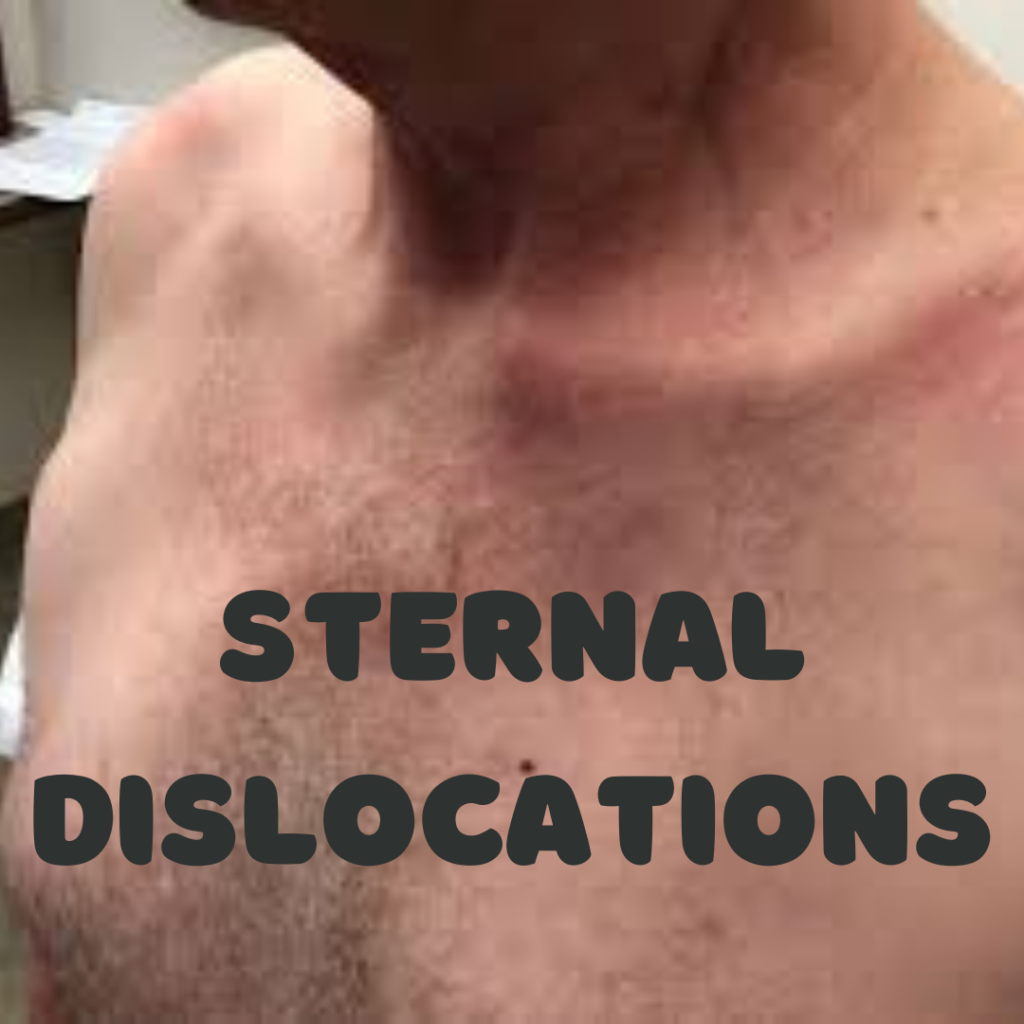
Understanding Sternal Dislocations
Sternal dislocations are relatively rare injuries characterized by the displacement of the sternum, or breastbone, from its normal position. This injury predominantly occurs due to trauma, which can result from high-impact situations such as motor vehicle accidents, falls, or sports-related incidents. Understanding the nature of sternal dislocations is vital for healthcare professionals, as timely recognition and appropriate management are crucial in preventing long-term complications.
The sternum serves as a central component of the thoracic skeleton, connecting the rib cage and providing structural support to the chest. It is composed of three parts: the manubrium, the body, and the xiphoid process. This flat bone plays a significant role in protecting vital organs, including the heart and lungs. When a dislocation occurs, it may involve a complete or partial separation of the sternum from the clavicular joint, complicating the stability of the rib cage and posing potential risks to surrounding structures.
In clinical practice, healthcare providers must recognize the mechanisms leading to sternal dislocations. Factors influencing the severity of this injury can include the angle of impact and the individual’s anatomy, such as pre-existing conditions or variations in rib structure. It is essential for practitioners to conduct thorough assessments and utilize imaging techniques, such as X-rays or CT scans, to ascertain the degree of dislocation and associated injuries, which may include damage to the underlying soft tissues. Proper evaluation and management strategies subsequent to diagnosis play a pivotal role in enhancing patient outcomes.
As medical practitioners continue to improve their understanding of sternal dislocations, it becomes increasingly clear that a focused approach to both recognition and treatment can significantly impact recovery and overall patient health.
Anatomy of the Sternum and Its Function
The sternum, also known as the breastbone, is a flat, elongated bone located in the center of the chest. It plays a crucial role in the structure of the thoracic skeleton. The sternum is comprised of three primary parts: the manubrium, the body, and the xiphoid process. Each component contributes to the overall stability and functionality of the ribcage.
The manubrium is the uppermost segment of the sternum and is shaped somewhat like a shield. It articulates with the clavicles (collarbones) at the sternoclavicular joint and serves as a point of attachment for several ribs via costal cartilages, helping to form the anterior portion of the rib cage. Below the manubrium lies the body of the sternum, which is the longest part. It connects to the second through the seventh pairs of ribs, thereby providing a strong framework that encases and protects the thoracic organs.
The xiphoid process is the smallest and lowest part of the sternum, initially cartilaginous in infants but typically ossifying into bone during adulthood. Though it is not directly involved in rib attachment, the xiphoid process serves as an important muscle attachment site, facilitating respiratory mechanisms through the diaphragm and abdominal muscles.
One of the primary functions of the sternum is to safeguard vital organs such as the heart and lungs, which reside within the thoracic cavity. By providing structural support and an anchor point for the ribcage, the sternum helps maintain the integrity of the chest wall throughout the respiratory process, allowing for efficient inhalation and exhalation.
The sternum’s anatomical design not only underscores its function in respiratory health but also highlights its importance in overall thoracic mechanics. Understanding the anatomy of the sternum is essential for recognizing how injuries or conditions affecting this structure, such as sternal dislocations, can impact respiratory function and overall health.
Causes and Mechanisms of Sternal Dislocations
Sternal dislocations are relatively uncommon injuries that typically result from significant external forces. The primary causes of these dislocations include trauma from high-impact situations such as car accidents, falls from significant heights, and sports-related injuries. These incidents can impart sufficient force onto the thoracic region, leading to an injury that dislodges the sternum from its normal articulation with the ribs and clavicles. Such injuries can particularly occur during severe blunt trauma where the upper body is subjected to a considerable impact.
Car accidents stand out as one of the most prevalent causes of sternal dislocations. The force generated during a collision often results in abrupt deceleration, which can cause the chest to collide with the steering wheel or seatbelt, risking injury to the sternum. Similarly, individuals involved in contact sports like football or rugby may experience sternal dislocations due to direct impacts or falls onto a rigid surface. In these scenarios, the violent motion can lead to maladaptive stress on the sternum and surrounding structures, culminating in dislocation.
Beyond external trauma, various risk factors may predispose individuals to sternal dislocations. Age is a significant determinant, as older adults generally possess a higher likelihood of sustaining injuries due to a potential decrease in bone density and strength. Individuals with underlying health conditions, such as osteoporosis or connective tissue disorders, may also be at greater risk. These conditions weaken the integrity of the thoracic skeleton, reducing its ability to withstand forces that would not normally result in dislocation in a healthier population. Understanding these mechanisms and risk factors is crucial for prevention and timely intervention in managing sternal dislocations.

Symptoms and Signs of Sternal Dislocation
Sternal dislocation, a relatively rare occurrence, presents a specific set of symptoms and physical signs that aid in its identification. The most prominent symptom experienced by individuals with this condition is severe pain localized at the center of the chest. This pain often intensifies with movement, particularly when engaging in activities that require the upper body to exert force. Patients may describe the pain as sharp or stabbing, which can often be misattributed to other thoracic injuries, making an accurate diagnosis essential.
In addition to pain, swelling in the area around the sternum is commonly observed. This localized swelling may indicate inflammation and should prompt further evaluation by healthcare professionals. Bruising or discoloration of the skin may also occur, manifesting as a result of underlying tissue trauma associated with the dislocation. These visible signs, coupled with the pain experienced by the patient, are crucial for differentiating sternal dislocations from other injuries affecting the thoracic region, such as rib fractures or costochondral separations, which may present similar initial symptoms.
It is important to note that individuals suffering a sternal dislocation may experience limitations in movement. Activities that require raising the arms or taking deep breaths can induce pain and discomfort, leading to a guarded posture and reduced mobility. This limitation can significantly affect the patient’s day-to-day activities and quality of life. Recognizing these symptoms early is vital for prompt and appropriate treatment. Furthermore, understanding the nuances of these signs enables healthcare practitioners to distinguish sternal dislocations from alternative thoracic injuries effectively, ensuring that patients receive the correct diagnosis and care necessary for optimal recovery.
Diagnosis of Sternal Dislocations
The diagnosis of sternal dislocations is a critical step in managing this injury effectively. It typically begins with a comprehensive physical examination conducted by a healthcare provider. During this assessment, the clinician evaluates the patient’s symptoms, inquires about the mechanism of injury, and inspects any visible deformities or bruising in the area of the sternum. Palpation may reveal tenderness or abnormal movement, aiding in assessing the injury’s severity. Nonetheless, the initial examination alone often cannot confirm a diagnosis; therefore, imaging techniques are essential.
X-rays are commonly the first imaging modality used in the evaluation of suspected sternal dislocations. They are widely available and can quickly reveal obvious fractures or significant dislocation. However, standard X-rays may not provide sufficient detail regarding the injury’s extent or potential complications. In such cases, more advanced imaging is warranted to gain a clearer understanding of the dislocation.
Computed tomography (CT) scans represent a more sophisticated option for visualizing the sternum and surrounding structures. CT imaging offers higher-resolution images and three-dimensional reconstructions, providing clinicians with a more accurate depiction of the dislocation and potential associated injuries, such as damage to adjacent vascular or lung structures. For patients where additional soft tissue evaluation is critical, magnetic resonance imaging (MRI) may be utilized to assess the ligaments and cartilage surrounding the sternum.
In conclusion, the diagnostic process for sternal dislocations relies on a combination of thorough physical examination and advanced imaging techniques. This integrated approach allows healthcare providers to accurately determine the presence and severity of the dislocation, thus enabling the development of an appropriate treatment plan tailored to the patient’s individual needs. The choice of diagnostic modality depends on the specifics of the injury and the patient’s clinical situation, emphasizing the importance of a careful evaluation by healthcare professionals.
Treatment Options for Sternal Dislocations
Sternal dislocations, although relatively rare, require prompt and effective treatment to ensure proper healing and minimize complications. The treatment options range from conservative management to surgical interventions, depending on the severity of the dislocation and the patient’s overall health condition.
Initially, conservative management is often the first approach for treating sternal dislocations. This method is particularly favored for patients with stable dislocations. Rest and activity modification are crucial components of conservative treatment, allowing the affected area to heal without the pressures of regular activity. Additionally, physical therapy may be introduced to improve movement, enhance strength, and promote function. Therapeutic exercises can be tailored specifically to the patient’s needs, aiding in the recovery process while ensuring the patient remains as active as possible.
In some cases, however, conservative methods may not yield the desired results, especially if the dislocation is deemed unstable or if there are associated injuries. In such situations, surgical intervention may be warranted. Surgical options typically involve realignment and stabilization of the sternum, often using plates or wires to secure the dislocated bone to its proper position. The decision to proceed with surgery is influenced by various factors, including the patient’s age, overall health, and the presence of any additional injuries. A thorough diagnostic assessment is crucial to establishing the most appropriate course of treatment.
Post-treatment rehabilitation plays an essential role in recovery. Whether conservative or surgical, a structured rehabilitation program can facilitate the healing process, allow for gradual return to daily activities, and help the patient regain strength and flexibility. It is imperative for patients to adhere to their healthcare provider’s recommendations throughout their recovery journey to optimize outcomes and enhance their quality of life.

Rehabilitation and Recovery Process
The rehabilitation process following a sternal dislocation is critical for regaining mobility and strength while ensuring proper healing. Initially, medical professionals will guide patients through a structured recovery plan tailored to their specific condition. The primary goal during this phase is to minimize pain and inflammation while promoting healing in the affected area.
Physical therapy plays a vital role in the recovery journey. A physical therapist will design a personalized exercise program, focusing on gentle stretches and strength-training activities to enhance the range of motion. These exercises are typically initiated once the acute pain subsides, usually several weeks post-injury. Patients are advised to remain consistent with their therapy sessions to reinforce the healing process and prevent stiffness in the chest area.
In addition to professional therapy, various self-care strategies can be implemented at home to facilitate recovery. It is paramount for individuals to prioritize rest, allowing their bodies ample time to recuperate. Adequate hydration and a balanced diet rich in vitamins and minerals can also bolster recovery, supporting the body’s healing mechanisms.
Pain management during the rehabilitation phase is crucial. Patients may be prescribed pain relief medication or advised to use over-the-counter options to alleviate discomfort. Techniques such as cold compresses or heat applications can provide additional relief as well. Additionally, patients are encouraged to pay attention to body signals and avoid activities that exacerbate pain or discomfort.
As individuals progress, the gradual return to normal activities should be approached with care. It is essential to consult with healthcare providers prior to resuming any rigorous physical activity or sports. By adhering to a structured rehabilitation plan and maintaining open communication with medical professionals, individuals can optimize their recovery from a sternal dislocation effectively.
Potential Complications of Sternal Dislocations
Sternal dislocations, while not extremely common, can lead to significant complications if left untreated or improperly managed. One of the primary concerns associated with sternal dislocations is the potential for long-term pain. Many patients may experience chronic discomfort in the chest area due to the misalignment of the sternum, which can interfere with daily activities and diminish overall quality of life. This pain can manifest as sharp, localized sensations or a more generalized discomfort that becomes exacerbated by physical activities such as lifting or exercising.
Another serious implication of untreated sternal dislocations is dysfunction of the thoracic region. The sternum plays a crucial role in protecting vital organs located beneath it, including the heart and lungs. A dislocated sternum can result in compromised function of these organs, leading to complications such as difficulty in breathing or cardiac disturbances. Over time, this dysfunction may become more pronounced, leading to further health ramifications that complicate recovery efforts.
Furthermore, disregarded sternal dislocations can cause injuries to underlying organs, particularly in cases where the dislocation results from traumatic forces. Blunt chest trauma may lead to contusions, lacerations, or even punctures of surrounding structures, necessitating prompt medical intervention. Neglecting to address these injuries may result in severe complications, including internal bleeding, respiratory distress, or even life-threatening conditions.
Thus, it is imperative to recognize the significance of prompt treatment and ongoing follow-up care for individuals diagnosed with sternal dislocations. Early intervention can substantially minimize the risk of long-term pain, dysfunction, and serious organ injury, ultimately facilitating a more successful recovery and better overall health outcomes.
FAQ About Sternal Dislocations
Sternal dislocations, while relatively rare, often raise various concerns among patients and healthcare providers. Below, we address some of the most frequently asked questions associated with this type of injury.
How can one prevent sternal dislocations?
Preventing sternal dislocations typically involves minimizing risks associated with physical activities. Engaging in safe practices during sports, particularly contact sports, is crucial. Proper use of safety equipment, including chest protectors, can significantly reduce the risk of injury. Additionally, maintaining good physical fitness and muscle strength can help support ribcage stability and prevent falls. Consultation with sports professionals regarding techniques and safe practices is also advisable.
When should someone seek medical attention for a suspected sternal dislocation?
Immediate medical attention should be sought if an individual experiences significant chest pain, difficulty breathing, or any form of trauma to the chest area, particularly following an accident or a fall. Other concerning symptoms include swelling, bruising, or a visible deformity in the chest. Prompt evaluation by a healthcare professional is essential for accurate diagnosis and treatment.
What is the long-term outlook for recovery after a sternal dislocation?
The prognosis following a sternal dislocation depends on several factors, including the severity of the dislocation and the timeliness of treatment. Most individuals can expect a positive recovery, typically within a few weeks to months, especially with appropriate medical intervention. Physical therapy may be recommended to restore strength and mobility. While many patients return to their normal activities, some may encounter lingering discomfort or decreased chest wall mobility.
Can sternal dislocations heal without surgery?
Yes, many sternal dislocations can heal without surgical intervention, especially if the dislocation is mild and does not cause significant instability. Treatment typically includes rest, pain management, and sometimes physical therapy to regain strength and mobility. However, severe cases, particularly those involving fractures or breathing difficulties, may require surgical stabilization.
Are there any complications associated with sternal dislocations?
Yes, complications can arise depending on the severity of the dislocation. Potential complications include chronic chest pain, restricted mobility, and in rare cases, damage to nearby organs such as the heart or lungs. Inadequate healing may also lead to long-term discomfort or instability in the chest wall. Seeking prompt medical attention can help reduce the risk of complications.
These questions highlight the importance of awareness and education regarding sternal dislocations, aiding both prevention and management efforts effectively.

Discover more from HUMANITYUAPD
Subscribe to get the latest posts sent to your email.

